

The Four Bases Of Dna
Chapter nine: Introduction to Molecular Biology
9.1 The Construction of DNA
Learning Objectives
By the end of this section, yous will exist able to:
- Describe the structure of DNA
- Depict how eukaryotic and prokaryotic DNA is bundled in the cell
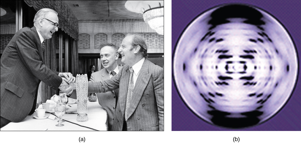
In the 1950s, Francis Crick and James Watson worked together at the University of Cambridge, England, to decide the structure of DNA. Other scientists, such as Linus Pauling and Maurice Wilkins, were also actively exploring this field. Pauling had discovered the secondary structure of proteins using Ten-ray crystallography. X-ray crystallography is a method for investigating molecular construction past observing the patterns formed past X-rays shot through a crystal of the substance. The patterns give important data about the structure of the molecule of involvement. In Wilkins' lab, researcher Rosalind Franklin was using 10-ray crystallography to understand the structure of Deoxyribonucleic acid. Watson and Crick were able to piece together the puzzle of the Dna molecule using Franklin's data (Figure 9.2). Watson and Crick likewise had central pieces of data available from other researchers such as Chargaff's rules. Chargaff had shown that of the four kinds of monomers (nucleotides) present in a DNA molecule, 2 types were always present in equal amounts and the remaining two types were too e'er present in equal amounts. This meant they were ever paired in some manner. In 1962, James Watson, Francis Crick, and Maurice Wilkins were awarded the Nobel Prize in Medicine for their work in determining the structure of Dna.
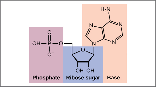
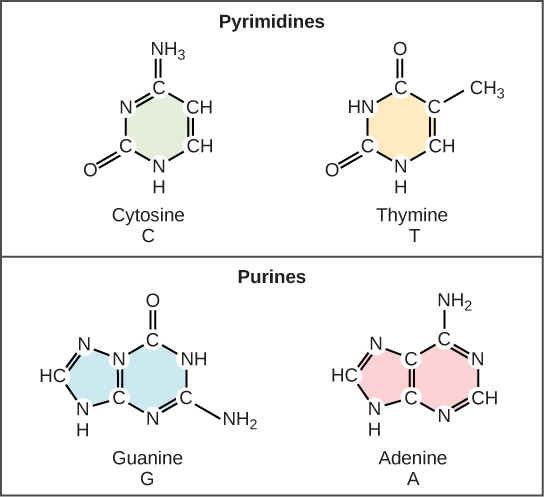
Now let'southward consider the structure of the 2 types of nucleic acids, dna (DNA) and ribonucleic acid (RNA). The edifice blocks of DNA are nucleotides, which are made up of three parts: a deoxyribose (5-carbon sugar), a phosphate group, and a nitrogenous base of operations (Effigy ix.three). In that location are four types of nitrogenous bases in Dna. Adenine (A) and guanine (G) are double-ringed purines, and cytosine (C) and thymine (T) are smaller, unmarried-ringed pyrimidines. The nucleotide is named according to the nitrogenous base it contains.
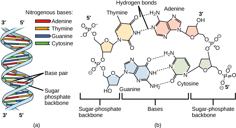
The phosphate grouping of one nucleotide bonds covalently with the saccharide molecule of the next nucleotide, and so on, forming a long polymer of nucleotide monomers. The sugar–phosphate groups line up in a "backbone" for each single strand of Dna, and the nucleotide bases stick out from this backbone. The carbon atoms of the 5-carbon sugar are numbered clockwise from the oxygen every bit i′, two′, three′, iv′, and 5′ (ane′ is read every bit "one prime"). The phosphate group is attached to the 5′ carbon of one nucleotide and the 3′ carbon of the next nucleotide. In its natural land, each DNA molecule is actually equanimous of two unmarried strands held together along their length with hydrogen bonds between the bases.
Watson and Crick proposed that the DNA is made up of two strands that are twisted effectually each other to form a correct-handed helix, chosen a double helix. Base-pairing takes identify between a purine and pyrimidine: namely, A pairs with T, and 1000 pairs with C. In other words, adenine and thymine are complementary base pairs, and cytosine and guanine are likewise complementary base pairs. This is the ground for Chargaff's rule; because of their complementarity, there is equally much adenine equally thymine in a DNA molecule and as much guanine as cytosine. Adenine and thymine are connected by two hydrogen bonds, and cytosine and guanine are connected by three hydrogen bonds. The ii strands are anti-parallel in nature; that is, one strand will have the three′ carbon of the sugar in the "upwardly" position, whereas the other strand will have the 5′ carbon in the upward position. The diameter of the Deoxyribonucleic acid double helix is uniform throughout because a purine (two rings) ever pairs with a pyrimidine (one ring) and their combined lengths are always equal. (Figure 9.4).
The Structure of RNA
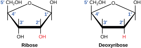
There is a second nucleic acrid in all cells called ribonucleic acrid, or RNA. Like DNA, RNA is a polymer of nucleotides. Each of the nucleotides in RNA is made up of a nitrogenous base, a 5-carbon sugar, and a phosphate group. In the case of RNA, the five-carbon sugar is ribose, not deoxyribose. Ribose has a hydroxyl grouping at the two′ carbon, unlike deoxyribose, which has merely a hydrogen atom (Figure 9.5).
RNA nucleotides contain the nitrogenous bases adenine, cytosine, and guanine. Notwithstanding, they do not contain thymine, which is instead replaced by uracil, symbolized by a "U." RNA exists as a unmarried-stranded molecule rather than a double-stranded helix. Molecular biologists have named several kinds of RNA on the footing of their function. These include messenger RNA (mRNA), transfer RNA (tRNA), and ribosomal RNA (rRNA)—molecules that are involved in the product of proteins from the DNA code.
How DNA Is Arranged in the Cell
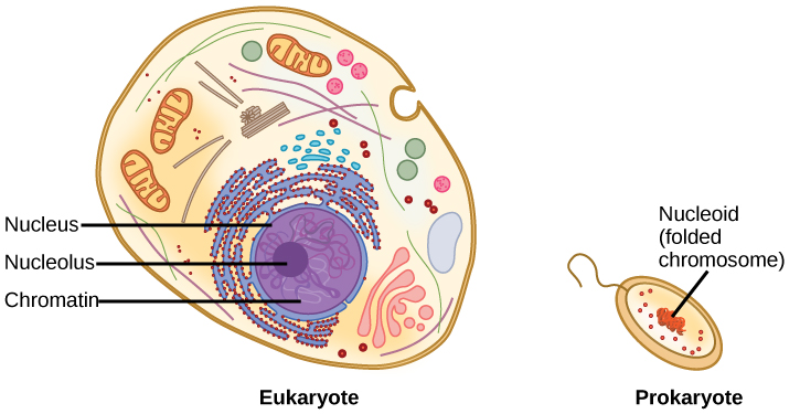
Dna is a working molecule; information technology must exist replicated when a cell is ready to split up, and information technology must be "read" to produce the molecules, such as proteins, to carry out the functions of the cell. For this reason, the Dna is protected and packaged in very specific means. In addition, DNA molecules tin be very long. Stretched end-to-finish, the Dna molecules in a single human jail cell would come to a length of about two meters. Thus, the DNA for a jail cell must exist packaged in a very ordered fashion to fit and part inside a structure (the cell) that is not visible to the naked heart. The chromosomes of prokaryotes are much simpler than those of eukaryotes in many of their features (Figure 9.six). Well-nigh prokaryotes contain a single, circular chromosome that is establish in an area in the cytoplasm called the nucleoid.
The size of the genome in one of the most well-studied prokaryotes, Escherichia coli, is iv.6 1000000 base pairs, which would extend a altitude of most 1.six mm if stretched out. Then how does this fit within a small bacterial jail cell? The DNA is twisted across the double helix in what is known as supercoiling. Some proteins are known to exist involved in the supercoiling; other proteins and enzymes help in maintaining the supercoiled structure.
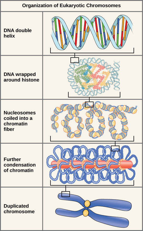
Eukaryotes, whose chromosomes each consist of a linear DNA molecule, use a different type of packing strategy to fit their DNA within the nucleus. At the most bones level, Dna is wrapped around proteins known as histones to grade structures chosen nucleosomes. The DNA is wrapped tightly around the histone core. This nucleosome is linked to the next one by a short strand of Dna that is free of histones. This is likewise known equally the "chaplet on a string" structure; the nucleosomes are the "chaplet" and the short lengths of DNA betwixt them are the "string." The nucleosomes, with their Dna coiled around them, stack compactly onto each other to course a 30-nm–wide cobweb. This cobweb is further coiled into a thicker and more than compact structure. At the metaphase stage of mitosis, when the chromosomes are lined upwardly in the center of the cell, the chromosomes are at their about compacted. They are approximately 700 nm in width, and are constitute in association with scaffold proteins.
In interphase, the phase of the cell cycle between mitoses at which the chromosomes are decondensed, eukaryotic chromosomes have two singled-out regions that can be distinguished by staining. There is a tightly packaged region that stains darkly, and a less dense region. The darkly staining regions normally contain genes that are not agile, and are found in the regions of the centromere and telomeres. The lightly staining regions commonly contain genes that are active, with Deoxyribonucleic acid packaged effectually nucleosomes only non further compacted.
Concept in Action

Watch this animation of DNA packaging.
Section Summary
The model of the double-helix construction of DNA was proposed past Watson and Crick. The DNA molecule is a polymer of nucleotides. Each nucleotide is composed of a nitrogenous base, a v-carbon sugar (deoxyribose), and a phosphate group. In that location are four nitrogenous bases in DNA, ii purines (adenine and guanine) and 2 pyrimidines (cytosine and thymine). A Dna molecule is composed of two strands. Each strand is composed of nucleotides bonded together covalently between the phosphate group of 1 and the deoxyribose sugar of the next. From this backbone extend the bases. The bases of one strand bail to the bases of the 2d strand with hydrogen bonds. Adenine always bonds with thymine, and cytosine always bonds with guanine. The bonding causes the two strands to spiral around each other in a shape called a double helix. Ribonucleic acid (RNA) is a 2d nucleic acrid found in cells. RNA is a unmarried-stranded polymer of nucleotides. It also differs from Deoxyribonucleic acid in that it contains the sugar ribose, rather than deoxyribose, and the nucleotide uracil rather than thymine. Various RNA molecules office in the procedure of forming proteins from the genetic lawmaking in DNA.
Prokaryotes contain a single, double-stranded round chromosome. Eukaryotes contain double-stranded linear DNA molecules packaged into chromosomes. The DNA helix is wrapped around proteins to form nucleosomes. The protein coils are farther coiled, and during mitosis and meiosis, the chromosomes become even more profoundly coiled to facilitate their movement. Chromosomes have ii distinct regions which can be distinguished by staining, reflecting dissimilar degrees of packaging and determined by whether the DNA in a region is beingness expressed (euchromatin) or non (heterochromatin).
Glossary
deoxyribose: a 5-carbon saccharide molecule with a hydrogen atom rather than a hydroxyl grouping in the 2′ position; the saccharide component of Deoxyribonucleic acid nucleotides
double helix: the molecular shape of Deoxyribonucleic acid in which 2 strands of nucleotides wind around each other in a spiral shape
nitrogenous base: a nitrogen-containing molecule that acts as a base of operations; often referring to one of the purine or pyrimidine components of nucleic acids
phosphate grouping: a molecular group consisting of a central phosphorus atom bound to four oxygen atoms

The Four Bases Of Dna,
Source: https://opentextbc.ca/biology/chapter/9-1-the-structure-of-dna/
Posted by: allenthwary.blogspot.com
0 Response to "The Four Bases Of Dna"
Post a Comment